
35-second body scan could tell you the sport you’re best suited to
By Candy Gibson
 HEALTH A 3D body scanner could replace manual anthropometric tests and MRIs for athletes.
 > Whole body scans for trauma patients cut time spent in emergency departments
Finding a world champion often falls to talent scouts and involves years of hard work but could be as simple as a 35-second body scan.
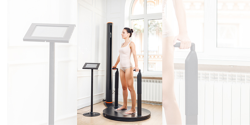
A new paper by UniSA sports scientist Professor Grant Tomkinson analyses how a $7500 3D portable whole-body scanner can identify sporting talent for particular codes and monitor body changes in athletes to ensure they are performing at their peak.
Prof Tomkinson, an adjunct researcher at UniSA based in the United States, has tested the reliability of a 3D whole-body scanner to measure athletes’ shape and composition, comparing it with X-rays, MRIs and manual tests, which involve bone calipers and girth tapes.
Anthropometry has been used for decades to identify talent, improve sports performance and health, but manual assessments and x-rays have their drawbacks, Prof Tomkinson says.
“3D scanning is less invasive than manual tests, and because it is fast, large samples can be easily measured, there is no need for physical contact, it doesn’t require a lot of expertise and it can measure body surface areas and volumes,” he says. “And, unlike x-rays, it doesn’t emit any potentially harmful radiation.”
Prof Tomkinson and a team from the University of North Dakota scanned 49 athletes (30 women and 19 men), subjecting them to a series of 35-second scans, extracting millions of measurements within 2mm accuracy.
All that is required is a 3D camera, a turntable and measurement extraction software.
“3D scans measure both cross-sectional areas and volumes and surface areas so they are generally better predictors of sporting success than manual tests,” he says.
In a few seconds a 3D body scan can measure the length of an arm and leg, the circumference of a thigh, or a body girth, as well as detect any asymmetries, such as scoliosis, or different leg lengths, which can affect the body’s musculature.
One of the main ways to select athletes for specific sports is to compare their body size and shape with the general population. For example, 3D body scans of elite rowers show that the best male rowers have much larger chests than the general population and elite female rowers have larger thighs than most women.
Similarly, marathon runners have a very defined physique – they are shorter, lighter, have longer legs relative to their torso and are leaner than the average person.
3D whole-body scanning has recently been used in several large anthropometric surveys of military personnel and the general population, but there is scant reliable anthropometric data on athletes beyond traditional manual measures, Prof Tomkinson says.
“Tracking changes in whole body and lean mass using 3D scanning would be an inexpensive and safer alternative to x-rays and magnetic resonance imaging (MRI) and could potentially be used to identify talent earlier and refine training to improve performance,” Prof Tomkinson says.
The research paper Reliability of the Styku 3D Whole-Body Scanner for the Assessment of Body Size in Athletes is published in the journal Measurement in Physical Education and Exercise Science.
Whole body scans for trauma patients cut time spent in emergency departments
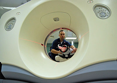 A patient undergoes whole body computed tomography (WBCT).
Taking whole body scans of emergency patients with traumatic injuries could lead to faster diagnosis and quicker treatment, according to research by a UniSA medical imaging student.
In a paper published in the European Journal of Radiology, UniSA student Elio Arruzza and his co-authors found that whole body computed tomography (WBCT) drastically cut the time spent in emergency departments as the procedure is far quicker than the traditional method of x-rays, ultrasound and selective CT scans of individual body regions.
WBCT is broadly defined as a CT scan of the head, cervical spine, chest, abdomen and pelvis.
The superior diagnostic accuracy of WBCT also makes it less likely that injuries are missed or even misdiagnosed, which happens with conventional imaging in up to 39 per cent of cases, Arruzza and his co-authors found.
However, WBCT imparts more radiation dose than other imaging procedures or non-WBCT, which clinicians, patients and families need to consider when weighing up the options.
In a meta-analysis of radiological procedures, Arruzza reviewed 14 studies comparing WBCT outcomes with conventional radiological procedures, or non-WBCT. While mortality rates, intensive care unit and hospital length stays were similar, time spent in emergency departments was significantly reduced.
“Our findings show that patients presenting with traumatic injuries can be diagnosed a lot faster with WBCT and therefore treated more quickly. This could in turn potentially reduce the impact of emergency department overcrowding, or ramping, which is a major problem in Adelaide and nationally,” Arruzza says.
“Much like the saying ‘time is brain’ in terms of stroke patients, ‘time is life’ for trauma patients. With expected improvements in the technology, we not only expect faster times but lower radiation doses as well.”
While the evidence suggests there is little difference in mortality rates between WBCT and non-WBCT, Arruzza says this is due to WBCT being reserved for critically ill patients.
“Less severely injured patients are still being scanned via the traditional x-ray, ultrasound and selective CT procedures so it is difficult to compare what their progress would be like if they received a full body scan,” he says.
More than five million people die from traumatic injuries each year, accounting for 9 per cent of global mortality and the leading cause of death among people under the age of 45.
About 60 per cent of trauma-related deaths occur within an hour after injury, compared to 30 per cent within 24 hours and the remainder over a longer period.
The research paper Systematic review and meta-analysis of whole-body computed tomography compared to conventional radiological procedures of trauma patients is authored by Elio Arruzza, Shayne Chau and Dr Janine Dizon.
Listen to Elio Arruzza explain the study in more detail in this interview with UniSA journalism lecturer Neelu Sharma.
Other Stories
- 35-second body scan could tell you the sport you’re best suited to
- Insects inspire infection resistant dental implants
- Is it a bird, a plane? Not superman, but a flapping wing drone
- Could our beloved Aussie rules footy be turning into soccer?
- From the Vice Chancellor
- Achievements and Announcements
- Health students step up to the plate to help curb Australia’s $170b medical bill
- Remote internships providing students with rewarding experiences
- Video: How to keep your brain healthy
- Which is more creative, the arts or the sciences?
- Local partnership delivers world-leading ‘smarts’ to shipbuilding
- MBA student creates Australia’s most eco-friendly magazine wrap
- Pandemic prompts students to solve challenges creatively
- The latest books from UniSA researchers
- In Pictures: Open August and VC Excellence Awards online event